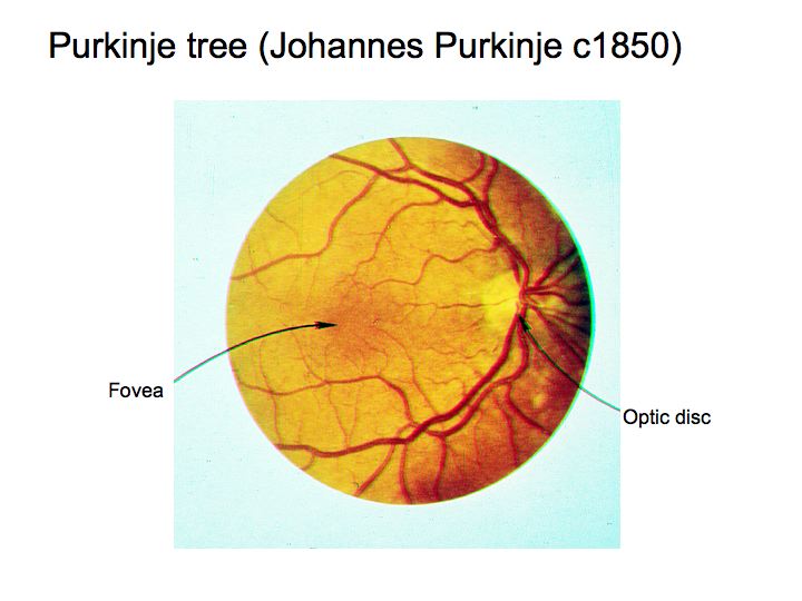
Apart from the neural tissue between the lens and the light receptors of your eye, there is also a bunch of tiny little blood vessels that branch all over the surface of the retina and provide it with food and nourishment. The pattern of blood vessels is called the Purkinje tree (after the Bohemian physiologist who first described it). The Purkinje tree branches out from the optic disk (blind spot) where the blood supply comes in and the optic nerve goes out. Notice also that there are few, if any, blood vessels in the the fovea, the center of your visual field.
Why don't you see the blood vessels when you look into somebody's eyes? If the light source is behind you, so that your head is between the light source and the eye you are studying, then your head will cast a shadow that interferes with the light from the point source arriving at your friend's eye. As a result, when you look in to measure the retinal image you see nothing beyond what is in your heart. If you move to the side of the light path, the image at the back of your friend's eye will be reflected towards the light source, following a reversible path. Since you are now on the side, out of the path of the light source, no light will be sent towards your eye.
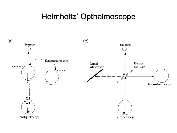
Helmholtz built the first ophthalmoscope. One needs to arrange a light path using half-silvered mirrors so that the examiner's eye does not cast a shadow. A bright source of light is required since the back of the human eye is not very reflective.
Stabilized vision: Why can't you see your own blood vessels in normal everyday vision? From what I've described to you, they are directly in the light path between the lens and the retina. So shouldn't you be seeing the shadow cast by these blood vessels on the surface of your retina? The reason you can't see thse blood vessels reveals a very important phenomenon about the way your visual system works. Notice that these blood vessels are fixed with respect to your retina. That is, as you move your eye about, since the blood vessels are attached to your retina, they move about with it, forming a fixed relationship. The pattern of retinal blood vessels does not change as you move your eye. Such a pattern - one that is fixed with respect to your retina even as the position of your eye changes - is called a stabilized image. The surprising fact about your eye is that it is insensitive to stabilized images.
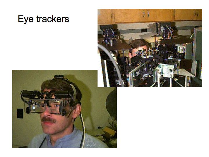
Special devices have been built that stabilize images. These devices are used for doing certain kinds of vision experiments that require careful control over eye movements. The device works by sensing the position of your eye. Each time you move your eye (even a little bit), it moves the visual stimulus by the same amount. In this way, the visual stimulus is projected onto your retina in such a way that even if you move your eyes the stimulus remains fixed with respect to your retina. Under such viewing conditions - called stabilized viewing conditions - the image of the world that is projected onto your retina does not make it to your conscious awareness. After a few seconds of stabilization, the entire scene seems to fade and disappear. The stabilized scene of the world disappears just like the shadows of your blood vessels disappear.
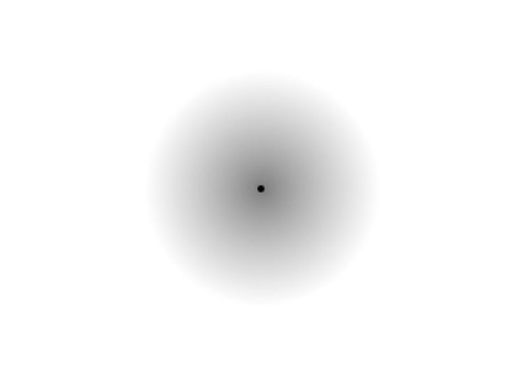
Stabilized image demo
One can't really demonstrate this in class without one of these expensive eye-trackers, but we can get close. Above is a small dot with a blurry gray circle around it. Since the transition is very gradual (from gray circle to white background), if you hold your eyes as steady as possible while fixating the central dot, the boundary of the blurry gray circle seems to disappear.
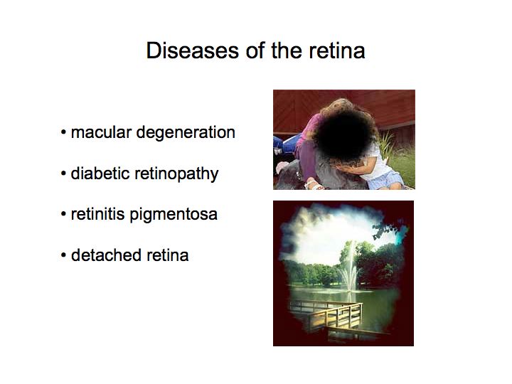
Diabetic retinopathy: swelling and abnormal growth of capillaries, insufficient oxygen supply to the retina, may cause bleeding into the vitreous. Treated by using a laser to seal off blood vessels.
Detached retina: the retina separates from the pigment epithelium, scotoma (blind spot) in the area that detaches. Caused by glaucoma or by injury. Use laser, heat, or cold probe to cause scar tissue to form and hold the retina in place.
Macular degeneration: degeneration of the part of the retina called the macula that includes the fovea and surrounding region. Leads to loss of central vision. The most common form is age related.
Retinitis pigmentosa: a hereditary disease, initially affects rods and peripheral vision, then gradually starts to affect cones and eventually leads to complete blindness.
Retinal implants: Because there are no good treatments for some retinal diseases (e.g., retinitis pigmentosa) there is interest in developing technology that would help alleviate these diseases. Some ideas involve redirecting the image to a healthy region of the retina. For example, one might have a mini video camera and a mini flat-panel display all mounted on eye glasses, with a computer to process the incoming images and special optics for projecting the processed images into the eye to a healthy region of the retina. This is not so far in the future.
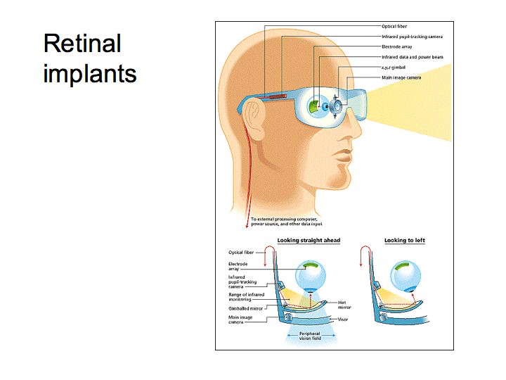
Conceptual design for a retinal implant
Other plans involve implanting a microchip with a digital camera on one side and stimulating electrodes on the other side, analogous to a cochlear implant. Obviously, it is very difficult to properly stimulate a large number of optic nerve fibers so it will be quite a number of years before this will be possible.
At present, only one vision-enhancement system that exploits advanced optoelectronics is on the market: the Low Vision Enhancement System (LVES), developed in the Wilmer Eye Institute at the Johns Hopkins University School of Medicine. This system provides variable focus and contrast rather than neuronal stimulation.
A company called Second Sight was founded in 1998 to create a retinal prosthesis to provide sight to patients blinded from retinal diseases. They have a prototype, experimental device that has been tested in a few individuals.
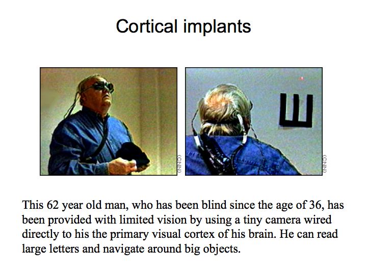
This 62 year old man, who has been blind since the age of 36, has been provided with limited vision by using a tiny camera wired directly to the primary visual cortex of his brain. The reports are that he can read large letters and navigate around big objects. He does not see an image. Rather, he perceives up to 100 specks of light that appear and disappear, like stars that come and go behind passing clouds, as his field of vision shifts.
Current research is aimed at extending the success of these experimental systems, to make them as successful and ubiquitous as cochlear implants. For example, there was a recent conference on Replacement Parts for the Brain. This conference brought together leading researchers throughout the country who focus on one of the newest frontiers of neuroscientific and bioengineering research: the intracranial implantation of computer chip models of brain function as neural prosthetics to replace damaged or dysfunctional brain tissue.
Further information about research and development of retinal implants.
Meanwhile, good electronic resources, links, and texts relating to technology and services for the blind can be found at The American Foundation for the Blind and at The Royal National Institute for the Blind.
The retina is a highly organized, layered structure. The information begins at the receptors and flows through a second layer of cells, called bipolar cells, and then continues through a third layer of cells, called ganglion cells. In addition, at the level of the receptor to bipolar connections, there are cells called horizontal cells that connect from receptors to other receptors. And at the level of interconnections between the bipolar cells and the ganglion cells there are cells called amacrine cells that take as their input signals from bipolar cells and feed their output back onto other bipolar cells.
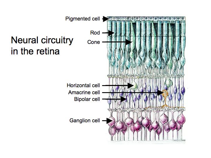
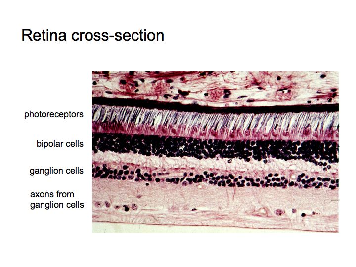
In the living eye, the neurons in the retina are all quite transparent so as not to interfere with the light on its way to the photoreceptors. In this picture, the neurons are stained so that you can see them. From bottom to top there are: clear ganglion cell axons that make up the optic nerve, stained ganglion cell bodies, bipolar cells, photoreceptors, and the black layer of pigment epithelium (special skin cells that absorb stray light and that help in the process of regenerating bleached photopigment).
None of the peripheral cells in the retina - the receptors, horizontal cells, bipolars, or amacrine cells - generate action poentials. Horizontal cells and amacrine cells don't even have proper axons. Rather these neurons respond with graded potentials. If the resulting current is large enough at their synapses, then they release neurotransmitter. It is only when you get to the level of the ganglion cells - with axons that reach a rather long distance into the brain - that you get action potentials.
A big theme in sensory neuroscience is that information is split apart and processed by separate subparts of the visual system. We have already seen a good example of parallel pathways: rods and cones. Typical properties of parallel pathways include:
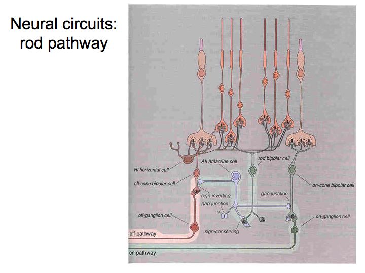
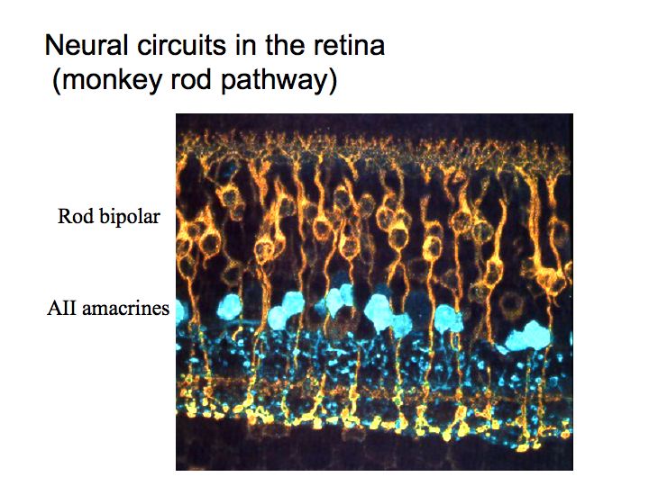
Midget, parasol, and bistratified retinal ganglion cells: Another example of parallel pathways. I'll discuss the midget and parasol cells here. The small bistratified retinal ganglion cells play a special role in color vision (see color lecture).
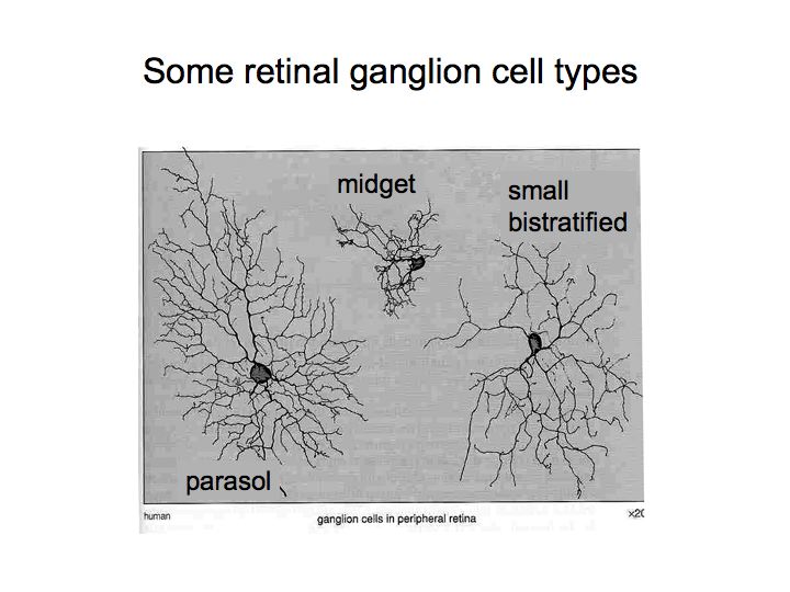
Anatomically distinct: Parasol dendritic trees are big, combine inputs from many cones. Midget trees are small. In the fovea, midget ganglion cells receive synapses from only one bipolar cell each which in turn receive synapses from only one cone. In the periphery, the dendritic fields of both ganglion cell types get bigger. It is easy to learn how to distinguish the two types. You need to pay attention to: (1) where you are in the retina, (2) how big is the tree.
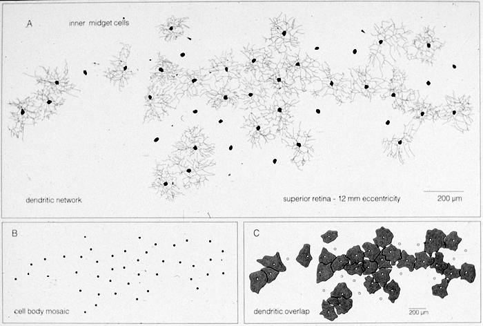
Complete coverage: A. Tracings of dendrites of 42 midget ganglion cells intracellularly stained. B. Cell body positions, regular distribution. C. Tracings around dendritic trees, perfect coverage with no overlap.
Recombine: Midget and parasol streams stay segregated in lateral geniculate nucleus (LGN), recombine to a large extent in visual cortex.
Physiologically distinct:
Parasol ganglion cells (magnocellular LGN cells):
Other parallel pathways initiated in the retina: There are about 40 ganglion cell types. About 90% of them project (send their axons to) the LGN and then on to visual cortex. The other 10% project to: superior colliculus in midbrain (involved in control of eye movements), pretectum in midbrain (pupil size, focus), superchiasmatic nucleus above optic chiasm (circadian rhythms).
Sound localization revisited (another example of a parallel pathway): We have seen an example of parallel pathways in audition as well. The two parts of the superior olive (MSO and LSO) are involved in sound localization. These pathways satisfy an analogous set of properties. They are functionally distinct: one extracts inter-aural timing differences (ITD) and the other extracts inter-aural intensity differences (IID). They are anatomically distinct: in 2 separate nuclei within the superior olive. There is complete coverage: in this case it's not coverage of the visual field, rather each nucleus has a full tonotopic map (covers all frequencies). They recombine: signals from MSO and LSO about ITD and IID information is combined later in the auditory pathways.
There are roughly a hundred million photoreceptor cells in each eye. There are two types of photoreceptors, rods and cones, with different shapes. Rods are specialized for low light levels (night vision), and cones are for high light levels and color vision.
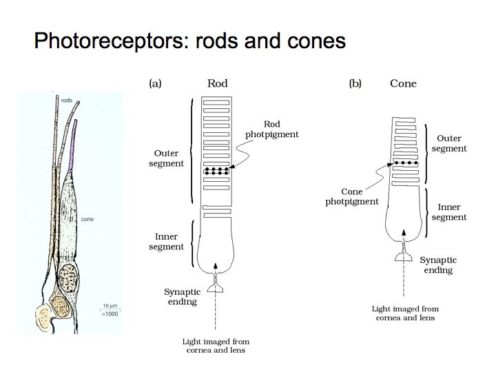
Electron microscope picture of some rods and cones followed by electron microscope picture (at higher magnification) of the folded membrane in the outer segment of a rod.
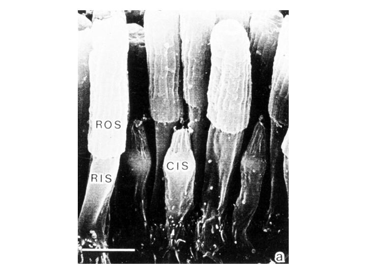
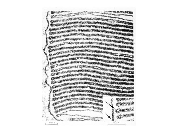
In this electron micrograph of a rod outer segment, you can see that the cellular membrane folds in and out. These internal membranes are highly structured and filled with photopigment molecules. In the rods, the photopigment is called rhodopsin and it is a complex molecule that is made up of two parts: one is called opsin and the other is a derivative of vitamin A.
When a quantum of light is absorbed by a molecule of rhodopsin, it changes the chemical state of the photopigment. The two parts of the molecule split. This change in state is called the isomerization of the photopigment. The isomerization sets off a biochemical chain reaction that eventually leads to an electrical current flowing in the rod. If the current is big enough, this neural signal is transmitted to the bipolar cells in the retina because neurotransmitter is released at the rod-bipolar synapse. Absorbing a single photon of light in a rod is enough to evoke a regular/reliable photocurrent. In fact, Hecht, Schlaer and Pirenne demonstrated psychophysically in the 40's that human subjects can reliably detect single photons.
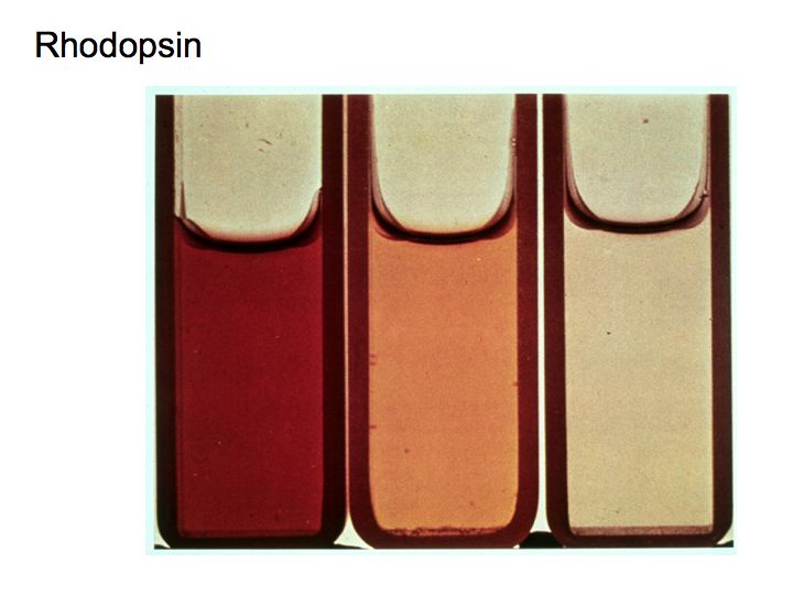
You can see the physical consequences of the isomerization in a chemistry lab. Each vial above contains a solution of rhodopsin that has been exposed to a different amount of light. The rhodopsin pigment changes color when exposed to light, called bleaching.
You can also see this phenomena directly in the living eye of certain animals (like cats and alligators) because the back of their eyes contains a white reflective surface, called the tapetum. That's why cats eyes appear to glow so brightly when they catch the beam of a car's headlight.
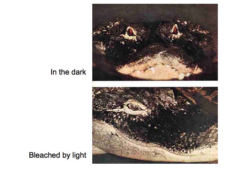
Alligator with red eyes; Alligator with no red eye
The above photographs show (1) what an alligator eye looks like after keeping in the dark for 24 hours so that the photopigment was fully regenerated and (2) what it looks like when exposed to light so that most of its rhodopsin is bleached.
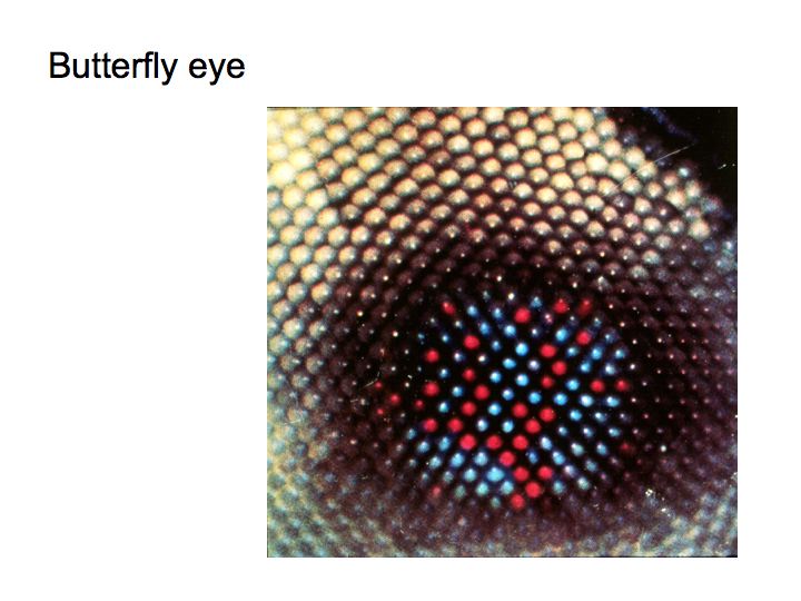
In the butterfly eye, you can see different colors corresponding to photoreceptors with different photopigments.
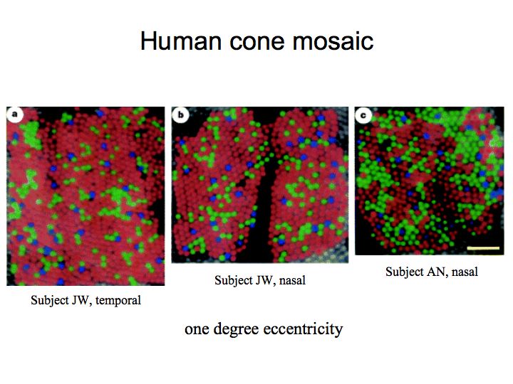
The rods all have the same photopigment, rhodopsin. But there are three different types of cones in the human retina, each with a slightly different photopigment. New optical techniques have made it possible to take pictures of the living human retina at very high magnification, and visualize the layout of the 3 different cone classes. The 3 photopigments absorb and reflect different wavelengths of light, giving rise to the different colors in the picture.
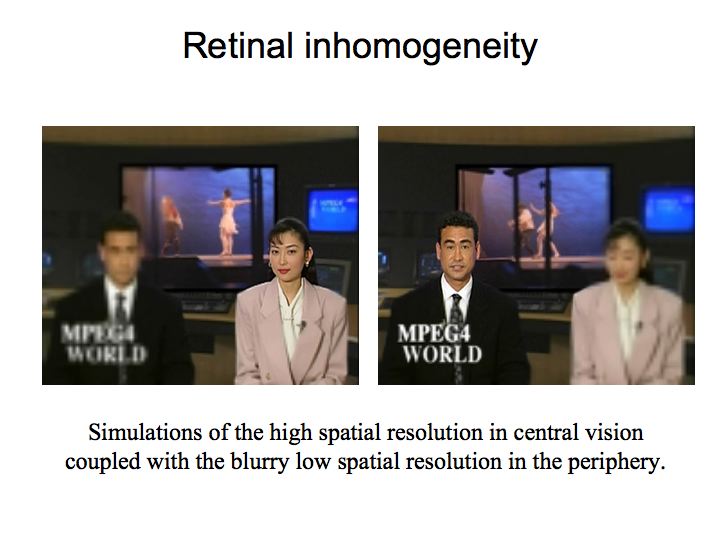
These two images are simulations of the high spatial resolution in central vision coupled with the blurry low spatial resolution in the periphery. The image on the left simulates fixating on the woman while the image on the right simulates fixating on the man.
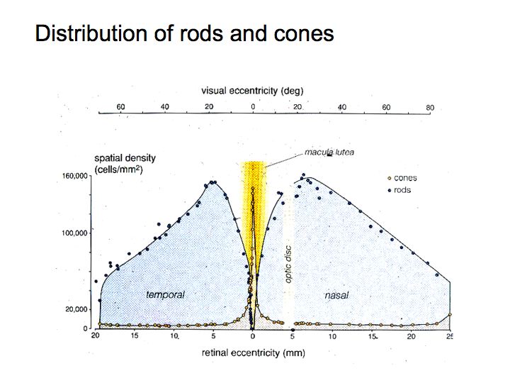
Rods and cones are not distributed uniformly across the retina. Most of the cones are in or near the fovea, the central part of the visual field. Most of the rods are in the peripheral part of the visual field. As mentioned above, rods are used for night vision and cones subserve daylight vision. Want to see a dim star on a moonless night? Cones are worthless. You need to use your rods. There are few rods in the center. So, you need to look slightly off to the side.
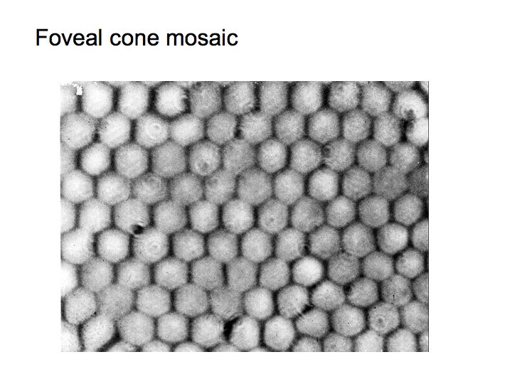
This pictures shows a cross-section of the foveal retina through the layer of the photoreceptor outer segments. The cones are packed very tightly, in nearly perfect hexagons, in the fovea.
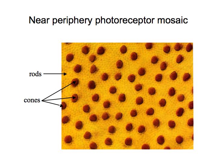
This picture shows a cross-section of the peripheral retina. The large brown circles are cones and the small yellow ones are rods. In the periphery, there are a lot more rods than cones, but the cones are big to absorb a lot of light. They are not packed in a perfect, orderly way as they are in the fovea.
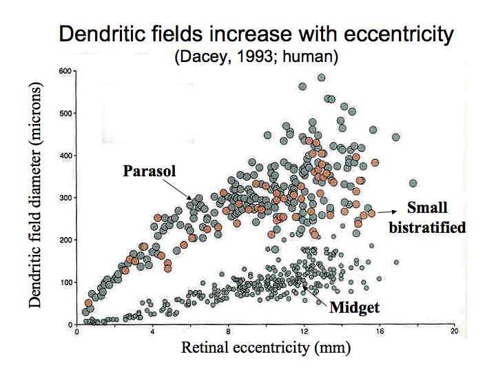
Peripheral ganglion cells pool inputs from many receptors, have bigger dendritic trees, have bigger receptive fields, and there are fewer of them. This allows you to have high spatial resolution for foveal viewing, but still have some vision in the periphery, and a small optic nerve.
One of the most important jobs (perhaps second only in importance to transduction by the photoreceptors) that the retina performs is light and dark adaptation. A fundamental challenge that is common to the signals carried by all visual neurons is that they must remain sensitive as the ambient light intensity varies over many orders of magnitude. There's well over a million-fold change in intensity between a starlit night and a bright sunny day at the beach. This is a challenge for the nervous system because neurons have a very limited response range: -80mV to +50 mV of graded potential in the non-spiking cells of the retina, or 0 to about 200 spikes per second for ganglion cells. The retina solves this problem by adapting to the ambient level of illumination. If the light level changes by a relatively small amount, then the visual system compensates for the change almost immediately. However, if the light level changes by a lot, then the eye takes a long time to re-adjust.
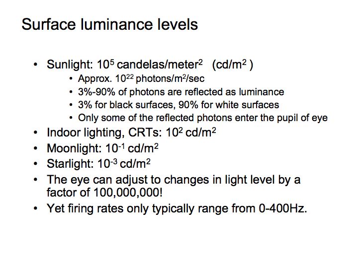
You must have noticed this phenomenon. When you are outside on a sunny day, and then walk into a darkened room, at first you can hardly see anything. For example, if people come into a darkened movie theater they are constantly tripping and stumbling over their seats on the way in. However, after being in the theater for a while, you adjust to the lower light level of the theater, and it becomes very easy to see your way around. This phenomenon is called adaptation, and the particular direction I just mentioned, where you adjust from the light to the dark, is called dark adaptation.


These figures shows the results of an experiment that quantifies this common experience. A subject was exposed to a bright adapting light, and then (while in the dark) their threshold was measured for detecting a very weak test light at various times after exposure to the bright light. The test light was violet in color. Filled symbols correspond to when the violet spot had threshold intensity and the violet color was evident at threshold. Open symbols mean that the violet color was not noticeable. Each curve corresponds to a different light level for the adapting light. The subject became more and more sensitive to the test light over time.
There are two very striking features. First, the data fall naturally into two parts - this double-branching is due to the transition from cones to rods. Second, the recovery is a fairly slow process - the subject's sensitivity to the light flash continued to increase over a very long time. In fact, after exposure to a very bright light, such as the subject was exposed to in the top curve of the figure, the subject did not fully recover to the best value for nearly forty minutes in the dark. During the initial portion of the curve, best vision is located at the fovea (right where you direct your eye), where cone density is highest. During the latter portion of the curve, best vision is in the parafovea (just to the side of where you direct your eye), where rod density is highest.
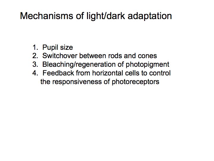
Light adaptation in the retina: The basic mechanisms responsible for controlling light/dark adaptation are quite complex. Part of the mechanism is the switch-over from rods to cones. But even within the pure rod (or pure cone) regime there is still significant adaptation. Only a small part of the adaptation is due to changes in pupil size; the pupil diameter only ranges from 1 or 2 mm to about 8 mm, for an increase in area (or total light entering the eye) of a factor of 16-64. A lot of the adaptation occurs in the photoreceptors themselves. Part of the adaptation within the photoreceptors is due to photopigment bleaching - less photopigment available at high light levels results in weaker responses to light increments at those high light levels. Adaptation within the photoreceptors is also helped along by feedback from horizontal cells onto the photoreceptors to control the responsiveness of the photoreceptors. If the horizontal cells respond strongly then they tell the photoreceptors to turn it down a bit.
Differences between rod and cone vision: There are a number of differences between daylight vision (photopic light levels, using your cones) and low-light levels (scotopic light levels, using rods). The first, higher branch of the above dark adaptation curves involves your photopic or cone system. Cones are most numerous at the fovea, and sensitivity is best there for photopic vision. Cones come in three types, responsible for your ability to discriminate colors (we'll talk about this more in the color lecture). Thus, in the above curves the violet color of the test light was evident for the photopic/cone portion of the curve. Photopic spectral sensitivity peaks at 560 nm, so that in photopic vision a yellow flash will be easiest to detect, and will appear the brightest. Cones are fast responders, so in the photopic range your ability to see fast flicker is high. Finally, cones are more sensitive to stimuli passing through the center of the pupil than passing through the edge of the pupil (this is called the Stiles-Crawford effect) because the cones are positioned and oriented toward the center of the pupil. For rods (scotopic vision, the second branch of the dark adaptation curves) we see different effects: sensitivity is highest in the parafovea (around the fovea), spectral sensitivity peaks at about 507 nm (so that green objects appear brightest), rods are slow (and can only detect very slow flicker) and show no Stiles-Crawford effect. At twilight light levels (mesopic light levels), both cones and rods are available for vision.
Afterimages: After being exposed to a bright light (e.g., flashbulb) you experience an afterimage, that is a (sometimes blueish) spot in your visual field. What causes the afterimage? The afterimage moves with you when you move your eyes. Why? After exposing a bit of your retina to bright light, the retinal becomes light adapted, but only where the light fell. In other words, light adaptation is local to the region of the retina that was stimulated. If after adapting to a small, bright light, you then look at a bright uniform field (e.g., a white wall), the adapted retina will be less sensitive, and that portion of the wall will look darker. This is called a negative afterimage. On the other hand, if you instead look at a very dark uniform field, there will again be an afterimage, but it will appear lighter than the background. This is called a positive afterimage, and the theory is that the adapted retina is signalling a weak stimulus when no stimulus is present, often referred to with the wonderful term: dark light.
Consequences for studying the rest of the visual system: Light/dark adaptation is a big effect and it happens at the front end of the visual system. One of the important consequences of this is that we need to be very clever to avoid effects of light/dark adaptation when we want to study later stages of visual processing. Let's say we are recording from a retinal ganglion cell. If we use visual stimuli that vary between being very bright and very dark, then we will see a lot of light adaptation (that is mostly due to processing in the photoreceptors and horizontal cells).
We can, however, safely ignore light adaptation by restricting our choice of visual stimuli to intensity distributions that modulate (transiently) about a fixed mean/background intensity. Examples are moving or flickering grating patterns and moving or briefly flashed bars that are either brighter or darker than the mean. The mean/background light intensity is typically chosen to be half the brightest intensity attainable on the display device.
Contrast: Contrast is the percent change in the light intensity in an image relative to the average light intensity.
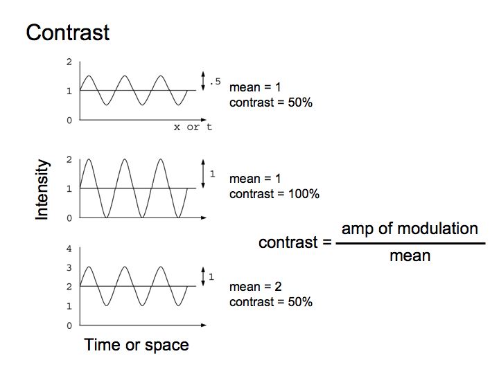
The contrast of a sinusoidal grating stimulus is defined as the difference between the highest and lowest intensities, divided by the sum of the two, or equivalently as the amplitude of modulation divided by the mean. The maximum possible contrast is 1 (100%), which is attained when the lowest intensity is zero and the highest intensity is twice the mean/background intensity. The contrast can go no higher than this, as this would imply having the intensity of the darkest portions of the stimulus go negative, which is physically impossible. The contrast of a stimulus is changed by scaling the image intensities above and below the fixed mean/background intensity. Thus, if we increase the highest and lowest light levels by, say, a factor of two, then mean light level will also increase, but contrast will remain unchanged. Light/dark adaptation in the retina results in a retina that responds to contrast rather than to absolute light levels.
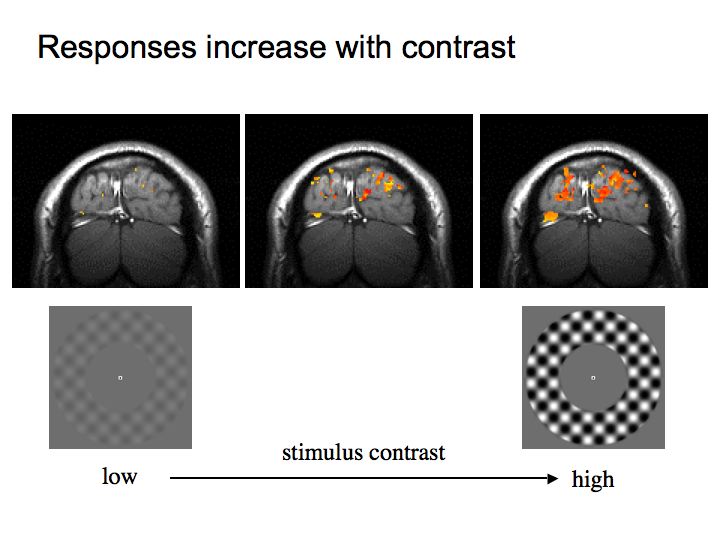
Neural responses in most of the visual system increase with contrast. The figure above, for example, shows that activity in visual cortex increase with contrast.
Summary: There are 4 mechanisms underlying light/dark adaptation:
It also has important consequences for how we design vision experiments - we often use transient stimuli like flickering grating patterns that are characterized in terms of their contrast.