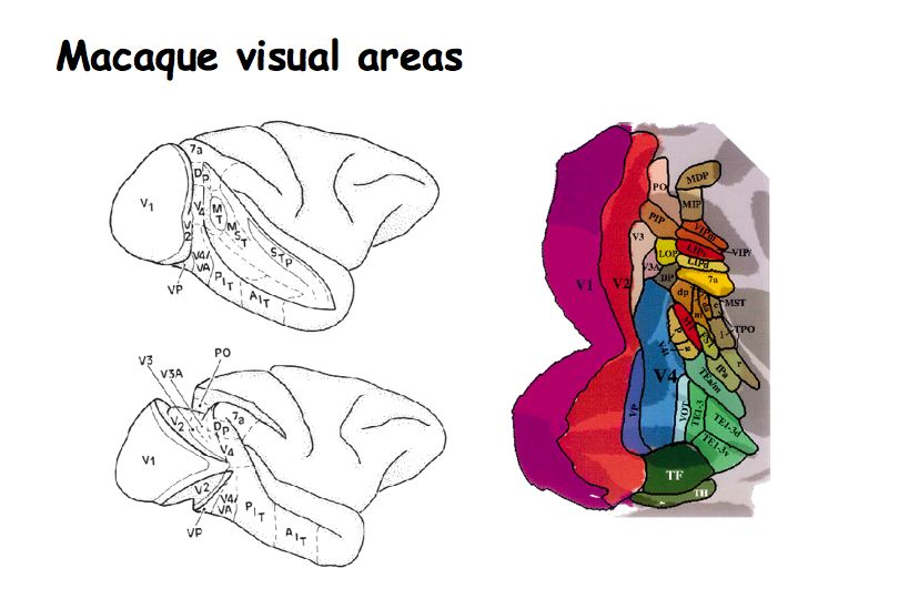
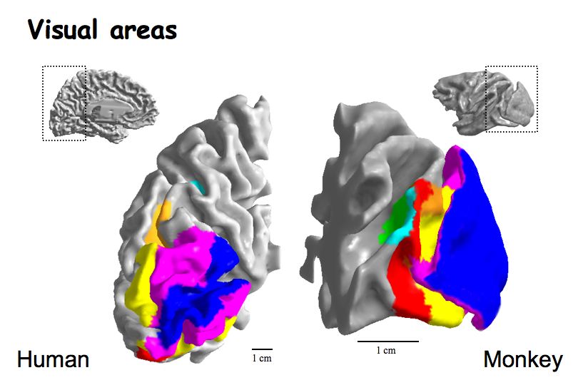
There are about 30 secondary visual areas after V1. Most lie in the occipital lobe. But some are in the parietal and temporal lobes as well. The diagram above of the monkey brain shows where the visual cortical areas are located and what they are called. Because the surface of the cortex is folded, you have to open up the sulci (left) or "flatten" the surface (right) to visualize the locations of the visual areas.
Brain flattening movie
In class we saw a video sequence showing a monkey brain physically being flattened. When we do fMRI experiments on humans, we also flatten their brains, but we do so computationally and render computer graphics pictures of their flattened brains.
Functional specialization hypothesis. This is the main hypothesis driving research into secondary visual cortical areas, specific brain area are involved in specific visual functions. Examples: MT is specialized for visual motion perception. IT is specialized for recognition. V4 is specialized for color vision.
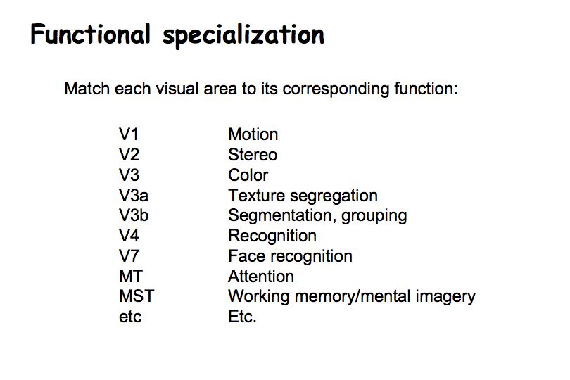
To date, however, there are very few examples in which we have successfully assigned one and only one function to a particular visual area. I have no doubt that each area performs a different function but we don't know yet what those functions are.
Defining visual cortical areas. The visual cortical areas are defined and identified based on both physiology and anatomy (the acronym we'll use is "PhACT").
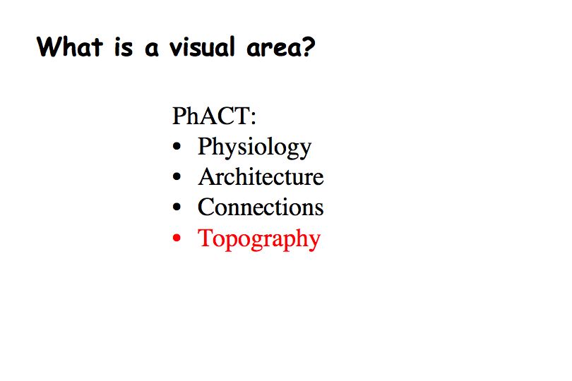
Physiology. Neurons in different brain areas are selective for different things (e.g., motion in MT).
Architecture. Cytoarchitecture (cell size, cell density, number of layers, density of axons, etc.) revealed through various different stains can change from one visual area to the next, helping to identify the boundaries between distinct brain regions.
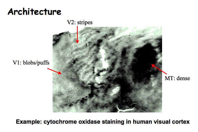
Cytochrome oxidase is an enzyme used in metabolism. Staining for it gives characteristically different patterns in different visual areas. In V1 it shows puffs or blobs. In V2 it yields a striped pattern. In V3 it yields a dense strip. In MT it yields a dense region.
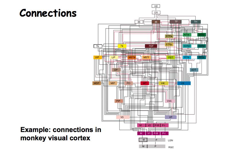
Connections. An anatomist can use chemicals that are transported by neurons (called tracers). The chemical is injected into one brain area. One then waits a while, then sacrifices the animal and stains for the tracer to see where it ends up. Some tracers are picked up by cell bodies and are transported to regions the cells project to; other tracers are picked up by axon terminals and are transported back to the corresponding cell bodies.
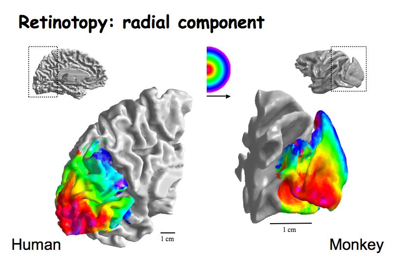
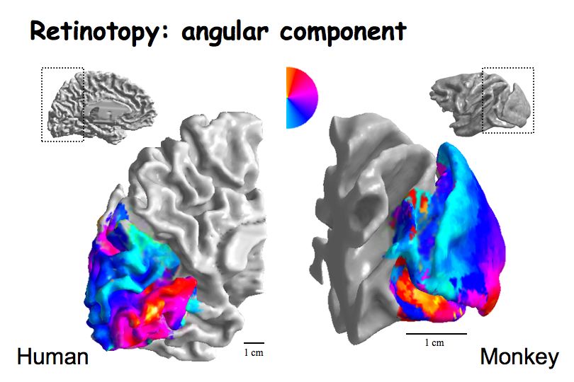
Topography. Each of the separate early visual areas contains a retinotopically organized map of the visual field. One can measure retinotopy using fMRI in the human brain using stimuli that traverse the visual field. The separate retinotopic maps define boundaries between visual areas. The boundaries between visual areas are marked by "reversals" in the retinotopic map.
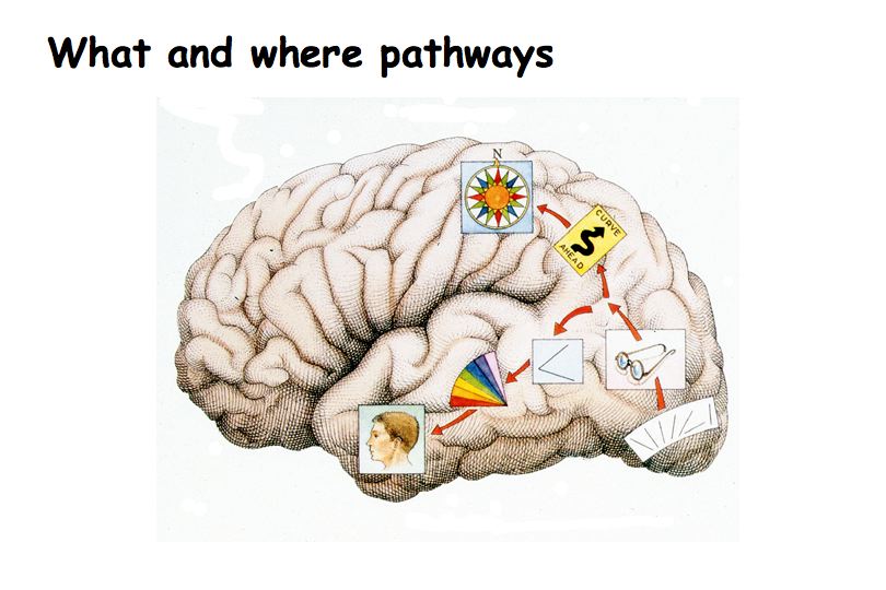
There are two distinct streams (parallel pathways). One goes to the parietal lobe and the other goes to the temporal lobe.
What do these two pathways do? Clinical observations have provided us with most of the information on this. One finds different deficits in patients who have lesions in these two different areas. The deficits are very different: