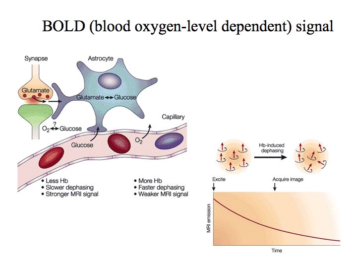
Physics of fMRI. The fundamental signal for blood oxygen level dependent (BOLD) functional magnetic resonance imaging (fMRI) comes from hydrogen atoms, which are abundant in the water molecules of the brain. In the presence of a magnetic field, these hydrogen atoms absorb energy that is applied at a characteristic radio frequency (~64 MHz for a standard, clinical 1.5 Tesla MRI scanner). After this step of applying radio-frequency excitation, the hydrogen atoms emit energy at the same radio frequency until they gradually return to their equilibrium state. The MRI scanner measures the sum total of the emitted radio-frequency energy. The measured radio-frequency signal decays over time, owing to various factors, including the presence of inhomogeneities in the magnetic field. Greater inhomogeneity results in decreased image intensity, because each hydrogen atom experiences a slightly different magnetic field strength, and after a short time has passed (commonly called T2*), their radio-frequency emissions cancel one another out. Deoxyhemoglobin introduces an inhomogeneity into the nearby magnetic field,whereas oxyhemoglobin does not. Hence, an increase in the concentration of deoxyhemoglobin causes a decrease in image intensity, and a decrease in deoxyhemoglobin causes an increase in image intensity.
Physiology of fMRI. The brain increases the local blood flow in reaction to the demand for glucose and oxygen. The details of this process are not fully understood but one theory posits that blood flow follows directly from increased synaptic activity. Astrocytes (specialize glial cells, not neurons, in the brain) surround both synapses and capillaries, and they are responsible for neurotransmitter recycling (taking the neurotransmitter out of the synapse quickly to stop its action on the post-synaptic membrane, causing a chemical change in the neurotransmitter molecules to deactivate them, and handing them back to the nearby neurons for reuse). All this takes a lot of energy.