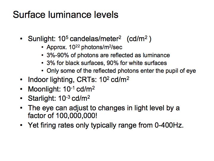
One of the most important jobs (perhaps second only in importance to transduction by the photoreceptors) that the retina performs is light and dark adaptation. A fundamental challenge that is common to the signals carried by all visual neurons is that they must remain sensitive as the ambient light intensity varies over many orders of magnitude. There's well over a million-fold change in intensity between a starlit night and a bright sunny day at the beach. This is a challenge for the nervous system because neurons have a very limited response range: -80mV to +50 mV of graded potential in the non-spiking cells of the retina, or 0 to about 200 spikes per second for ganglion cells. The retina solves this problem by adapting to the ambient level of illumination. If the light level changes by a relatively small amount, then the visual system compensates for the change almost immediately. However, if the light level changes by a lot, then the eye takes a long time to re-adjust.
You must have noticed this phenomenon. When you are outside on a sunny day, and then walk into a darkened room, at first you can hardly see anything. For example, if people come into a darkened movie theater they are constantly tripping and stumbling over their seats on the way in. However, after being in the theater for a while, you adjust to the lower light level of the theater, and it becomes very easy to see your way around. This phenomenon is called adaptation, and the particular direction I just mentioned, where you adjust from the light to the dark, is called dark adaptation.
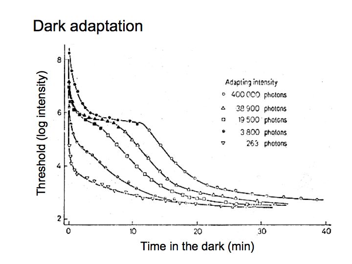
This figure shows the results of an experiment that quantifies this common experience. A subject was exposed to a bright adapting light, and then (while in the dark) their threshold was measured for detecting a very weak test light at various times after exposure to the bright light. The test light was violet in color. Filled symbols correspond to when the violet spot had threshold intensity and the violet color was evident at threshold. Open symbols mean that the violet color was not noticeable. Each curve corresponds to a different light level for the adapting light. The subject became more and more sensitive to the test light over time.
There are two very striking features. First, the data fall naturally into two parts - this double-branching is due to the transition from cones to rods. Second, the recovery is a fairly slow process - the subject's sensitivity to the light flash continued to increase over a very long time. In fact, after exposure to a very bright light, such as the subject was exposed to in the top curve of the figure, the subject did not fully recover to the best value for nearly forty minutes in the dark.
Light adaptation in the retina: The basic mechanisms responsible for controlling light/dark adaptation are quite complex. Part of the mechanism is the switch-over from rods to cones. But even within the pure rod (or pure cone) regime there is still significant adaptation. Only a small part of the adaptation is due to changes in pupil size; the pupil diameter only ranges from 1 or 2 mm to about 8 mm, for an increase in area (or total light entering the eye) of a factor of 16-64. A lot of the adaptation occurs in the photoreceptors themselves. Part of the adaptation within the photoreceptors is due to photopigment bleaching - less photopigment available at high light levels results in weaker responses to light increments at those high light levels. Adaptation within the photoreceptors is also helped along by feedback from horizontal cells onto the photoreceptors to control the responsiveness of the photoreceptors. If the horizontal cells respond strongly then they tell the photoreceptors to turn it down a bit.

Differences between rod and cone vision: There are a number of differences between daylight vision (photopic light levels, using your cones) and low-light levels (scotopic light levels, using rods). The first, higher branch of the above dark adaptation curves involves your photopic or cone system. Cones are most numerous at the fovea, and sensitivity is best there for photopic vision. Cones come in three types, responsible for your ability to discriminate colors (we'll talk about this more in the color lecture). Thus, in the above curves the violet color of the test light was evident for the photopic/cone portion of the curve. Photopic spectral sensitivity peaks at 560 nm, so that in photopic vision a yellow flash will be easiest to detect, and will appear the brightest. Cones are fast responders, so in the photopic range your ability to see fast flicker is high. Finally, cones are more sensitive to stimuli passing through the center of the pupil than passing through the edge of the pupil (this is called the Stiles-Crawford effect) because the cones are positioned and oriented toward the center of the pupil. For rods (scotopic vision, the second branch of the dark adaptation curves) we see different effects: sensitivity is highest in the parafovea (around the fovea), spectral sensitivity peaks at about 507 nm (so that green objects appear brightest), rods are slow (and can only detect very slow flicker) and show no Stiles-Crawford effect. At twilight light levels (mesopic light levels), both cones and rods are available for vision.
Afterimages: After being exposed to a bright light (e.g., flashbulb) you experience an afterimage, that is a (sometimes blueish) spot in your visual field. What causes the afterimage? The afterimage moves with you when you move your eyes. Why? After exposing a bit of your retina to bright light, the retinal becomes light adapted, but only where the light fell. In other words, light adaptation is local to the region of the retina that was stimulated. If after adapting to a small, bright light, you then look at a bright uniform field (e.g., a white wall), the adapted retina will be less sensitive, and that portion of the wall will look darker. This is called a negative afterimage. On the other hand, if you instead look at a very dark uniform field, there will again be an afterimage, but it will appear lighter than the background. This is called a positive afterimage, and the theory is that the adapted retina is signalling a weak stimulus when no stimulus is present, often referred to with the wonderful term: dark light.
Consequences for studying the rest of the visual system: Light/dark adaptation is a big effect and it happens at the front end of the visual system. One of the important consequences of this is that we need to be very clever to avoid effects of light/dark adaptation when we want to study later stages of visual processing. Let's say we are recording from a retinal ganglion cell. If we use visual stimuli that vary between being very bright and very dark, then we will see a lot of light adaptation (that is mostly due to processing in the photoreceptors and horizontal cells).
We can, however, safely ignore light adaptation by restricting our choice of visual stimuli to intensity distributions that modulate (transiently) about a fixed mean/background intensity. Examples are moving or flickering grating patterns and moving or briefly flashed bars that are either brighter or darker than the mean. The mean/background light intensity is typically chosen to be half the brightest intensity attainable on the display device.
Contrast: Contrast is the percent change in the light intensity in an image relative to the average light intensity.

The contrast of a sinusoidal grating stimulus is defined as the difference between the highest and lowest intensities, divided by the sum of the two, or equivalently as the amplitude of modulation divided by the mean. The maximum possible contrast is 1 (100%), which is attained when the lowest intensity is zero and the highest intensity is twice the mean/background intensity. The contrast can go no higher than this, as this would imply having the intensity of the darkest portions of the stimulus go negative, which is physically impossible. The contrast of a stimulus is changed by scaling the image intensities above and below the fixed mean/background intensity.
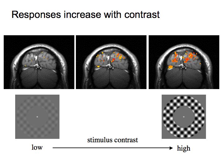
Neural responses in most of the visual system increase with contrast. The figure above, for example, shows that activity in visual cortex increase with contrast.
Summary: There are 4 mechanisms underlying light/dark adaptation:
It also has important consequences for how we design vision experiments - we often use transient stimuli like flickering grating patterns that are characterized in terms of their contrast.